
The Nobel Assembly at the Karolinska Institutet has awarded the 2026 Nobel Prize in Physiology or Medicine to Karl Deisseroth, Peter Hegemann, and Georg Nagel for their pioneering discoveries leading to optogenetics.
By harnessing light-gated ion channels—specifically channelrhodopsins derived from microalgae—and integrating them into mammalian cells, these researchers transformed how life scientists observe and control cellular machinery.
For molecular biologists, biotechnologists, and laboratory managers, optogenetics represents a technical triumph and a blueprint for high-precision experimental workflows.
The Molecular Mechanism: From Green Algae to Mammalian Membranes
The foundation of optogenetics began with an organism: Chlamydomonas reinhardtii, a single-celled green alga.
[470 nm Blue Light Flash]
         │
         ▼
┌───────────────────────────────── ┐
│ Channelrhodopsin-2 (ChR2)        │
│ Transmembrane Retinal Chromophore│
└───────────────────────────────── ┘
         │ (13-cis Photoisomerization)
         ▼
┌─────────────────────────────────┐
│ Pore Opening (~6 Å Diameter)    │
│ Passive Influx: Na+, K+, Ca2+   │
└─────────────────────────────────┘
         │
         ▼
[Rapid Membrane Depolarization (~1 ms)]
1. Biophysical Discovery (Hegemann & Nagel)
Biophysicists Peter Hegemann and Georg Nagel isolated and characterized Channelrhodopsin-1 (ChR1) and Channelrhodopsin-2 (ChR2). Unlike classical rhodopsins that rely on G-protein coupled cascades, channelrhodopsins are single-component, light-gated ion channels.
Upon absorption of a blue photon (~470 nm), the covalently bound retinal chromophore undergoes trans-to-cis isomerization, opening a cation-permeable pore within microseconds.
2. Genetic Integration & Bioengineering (Deisseroth)
Karl Deisseroth demonstrated that the ChR2 gene could be heterologously expressed in mammalian neurons using viral vectors (such as AAVs) driven by cell-type-specific promoters (e.g., Camk2a, Syn1).
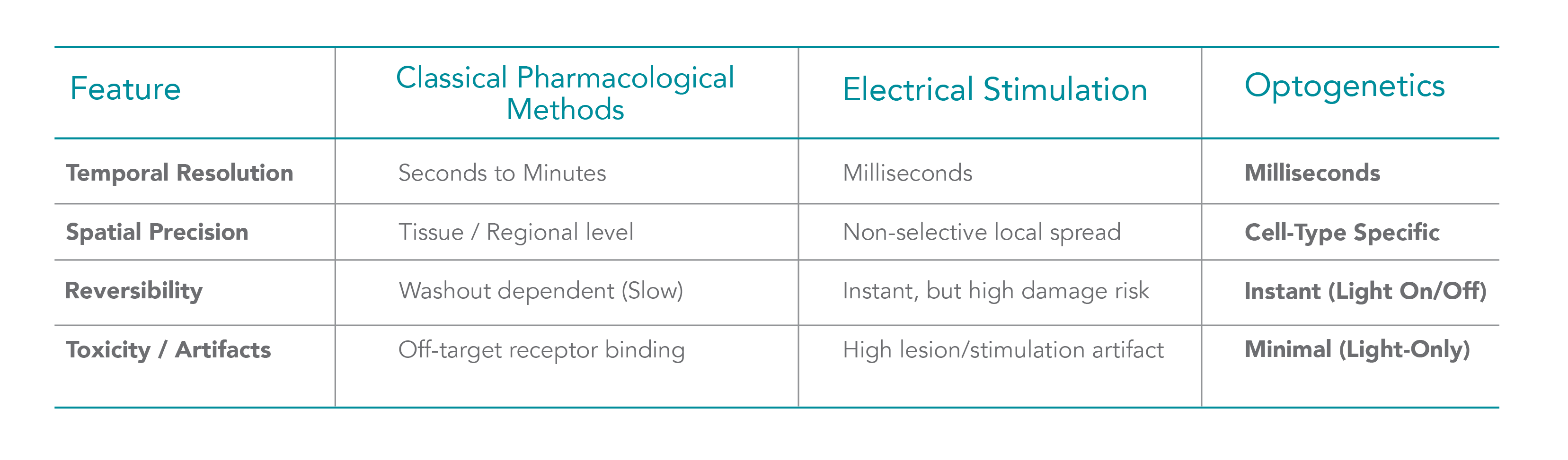
When exposed to targeted pulse sequences of blue light, these engineered cells depolarize on millisecond timescales, triggering action potentials with temporal precision.
Lab Operations & Workflow Optimization
Translating optogenetic protocols into reproducible experimental data requires tight control over biological reagents, vector delivery, and optoelectronic equipment.
Vector Construction & QC
-
Plasmid Design: Ensuring high-titer adeno-associated virus (AAV) or lentivirus preparations (typically > 10¹² GC/mL) with validated tissue-specific promoter sequences.
-
Transfection Control: Verifying reporter protein expression (e.g., ChR2-mCherry or ChR2-eYFP fusion constructs) via fluorescence microscopy prior to functional assays.
Light Delivery & Optical Hardware
-
Wavelength Integrity: Solid-state laser lines or narrow-band LEDs (470 nm for activation, 590 nm for halorhodopsin-mediated inhibition) with fast TTL modulation drivers.
-
Power Density Calibration: Maintaining precise irradiance at the tissue sample (typically 1–10 mW/mm²) to avoid thermal phototoxicity or unintended bleaching.
Media & Reagent Purity
-
Optogenetic channels rely on precise physiological balances of monovalent and divalent cations (Na⁺, K⁺, Ca²⁺). Standardized, ultra-pure buffer formulations and low-autofluorescence growth media are essential to maintain baseline membrane potentials and clear optical pathways.
Applications Across Biotechnology & Medicine
While initially developed for systems neuroscience, optogenetic principles are expanding across life science sectors:
-
Cardiomyocyte Research & Arrhythmia Models: Optogenetic control of cardiac tissue allows researchers to pace cultured cardiomyocytes without electrical contact artifacts, streamlining high-throughput cardiotoxicity screening.
-
Synthetic Biology & Gene Expression: Light-inducible transcriptional systems (e.g., photo-activatable Cre recombinase or Gal4/UAS systems) allow spatial patterning of gene expression in stem cell cultures and organoids.
-
Translational Therapeutics & Diagnostics: Clinical trials are actively using AAV-delivered channelrhodopsins to restore light sensitivity in retinal degenerative conditions such as Retinitis Pigmentosa.
Conclusion: A New Horizon for Precision Biology
The 2026 Nobel Prize highlights a fundamental evolution in life sciences: the transition from static, broad-spectrum interventions to dynamic, spatiotemporally controlled molecular tools. By demonstrating how a single algal protein can grant microsecond command over cell function, Deisseroth, Hegemann, and Nagel did more than solve a biophysical puzzle—they handed the scientific community a universal remote control for cellular mechanics. As these principles continue to inspire new methodologies in organoid research, cardiac modeling, and synthetic biology, optogenetics stands as a powerful reminder of how basic curiosity about simple organisms can ultimately reshape the landscape of modern medicine.